Electrical biology becomes useful when the question is precise
The word electrical is both scientifically important and easy to overextend. A heart rhythm tracing and a recording from scalp electrodes are real measurements of biological activity. Yet their existence does not establish that a person has one overall electrical balance that can be checked, drained or restored like a phone battery. To understand a recording, we need to ask where the signal comes from, how it was collected and which clinical decision it can inform.
I see precision here as an aid to curiosity. Naming the actual measurement makes electrical biology more interesting, not less. An ECG and an EEG can both produce lines on a screen, but a similar visual format does not make them equivalent. A map and a weather chart may both use colours and symbols while answering entirely different questions. Biological recordings deserve the same attention to purpose.
This article concerns electrocardiography and electroencephalography as clinical recordings. It does not teach interpretation of individual traces, which requires training and clinical context. Nor is it a recommendation for screening people without a clinical indication. The aim is to help a reader understand why a test is requested, prepare sensibly and discuss what its result actually changes.
Evidence: what an ECG records
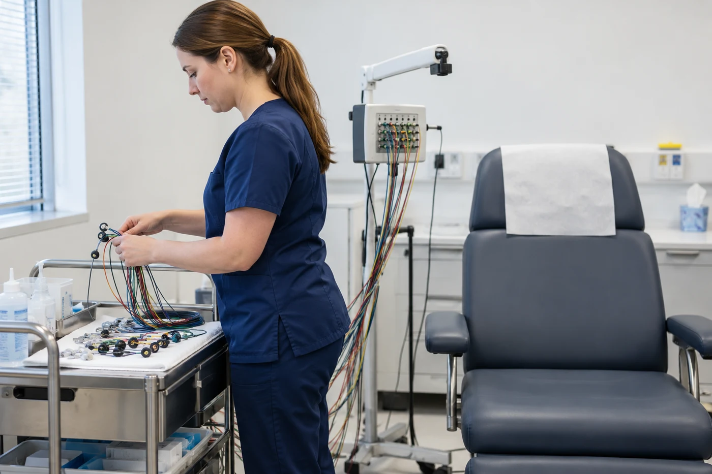
An electrocardiogram, abbreviated ECG or EKG, records electrical activity associated with the heart. The US National Heart, Lung, and Blood Institute explains that the recording can show heart rate, rhythm and the timing of electrical impulses. Electrodes placed on the skin connect to a recording machine. A standard recording does not deliver an electrical shock or recharge the heart [1].
The important distinction is between recording a signal and treating an organ. The small patches and leads are part of a measurement system. Their presence does not mean that electricity is being added to correct a supposed deficiency. A clinician interprets patterns in relation to symptoms, history, medicines and other information. The result becomes useful through that connection rather than through the mere presence of a trace.
The NHS describes resting, portable and exercise forms of ECG. Their settings differ because symptoms and clinical questions differ. A short resting recording samples one period. A portable device records over longer periods, while an exercise test examines a response under supervised conditions. Preparation and restrictions therefore depend on the specific test requested [2]. The correct question is which recording is appropriate, not which version seems most technologically impressive.
Evidence: what an EEG records
An electroencephalogram, or EEG, records electrical activity from the brain using electrodes on the scalp. The NHS identifies epilepsy as its main clinical use, with other applications in selected brain and sleep conditions. A trained professional attaches the electrodes, records activity and may ask the person to open their eyes or undertake other supervised tasks. The recording is later analysed by an appropriate specialist [3].
The National Institute of Neurological Disorders and Stroke describes EEG as monitoring brain electrical activity through the skull. It can be used in seizure assessment and in other defined neurological settings, including selected monitoring during altered consciousness or anaesthesia [4]. This is an observation of activity, rather than a photograph of brain structure. Different diagnostic techniques can therefore provide complementary information without one replacing all the others.
The recording also is not a reading of private thoughts, personality or moral character. Calling an EEG pattern a brain wave does not turn it into a complete account of a person's emotional life. The distinction matters when familiar scientific words are borrowed to support a much broader commercial claim. A measured signal must remain attached to the limits of the method that produced it.
Evidence: why the history can matter as much as the trace
NICE recommends a 12-lead ECG after a first suspected seizure to help identify cardiac conditions that can resemble an epileptic event. If the history and examination suggest epilepsy, an EEG may support diagnosis and help with classification. NICE explicitly advises that an EEG should not be used to exclude epilepsy [5]. The two tests can therefore appear in one investigation without being interchangeable.
A normal result is not a universal certificate that every relevant process is normal at every time. A recording is collected under particular circumstances and interpreted for a particular reason. When uncertainty remains, the clinician may recommend further assessment or a different recording. This does not mean the first test was pointless. A limited answer can still be useful when its limits are clearly explained.
A result discussion can also separate three decisions that people often combine: whether an event has been explained, whether further investigation is needed and whether treatment should begin. These are related decisions, but they are not identical. It is reasonable to ask which one the present test is meant to inform. If the answer is still uncertain, ask how that uncertainty will be managed rather than assuming that another test must automatically resolve it. This keeps the next step connected to an explicit purpose and gives the person a clearer account of the care plan.
Conversely, the word abnormal is not a complete diagnosis. Ask which finding is being described, how strongly it supports the proposed explanation and what alternative explanations remain. A report without an accompanying clinical discussion may feel definitive while leaving the central question unresolved. The sensible next step is interpretation with the referring team, rather than comparison with an unrelated trace found online.
Practical implications: prepare for the test you are actually having
For an EEG, the NHS advises clean, dry hair without styling products and following any additional instructions from the testing service. Different EEG formats can require different preparation. A sleep-deprived recording, for example, involves arrangements that are different from an ordinary appointment, including transport considerations [3]. Do not deliberately lose sleep merely because an online description mentions a sleep-deprived test.
Mayo Clinic advises taking usual medicines unless the care team says otherwise. It also explains that some supervised EEG procedures use flashing lights or changes in breathing to help assess activity, and that these can occasionally trigger a seizure in susceptible people. The team discusses risks and provides appropriate care [6]. Such procedures should not be copied at home to test whether a person reacts.
For ECG appointments, ask whether the test is resting, portable or exercise-based. The NHS notes practical preparation such as avoiding lotions on skin where electrodes will be attached and following the service's instructions for an exercise recording [2]. Tell the team about medicines and ask about any uncertainties. Self-directed changes to prescribed treatment can create a different problem without improving the usefulness of the test.
Practical implications: bring the event into the conversation
A clinician needs a description of the problem that prompted testing. Useful observations include what happened immediately before an episode, whether there was a warning, how long it lasted, what recovery was like and whether it has happened in other circumstances. A witness's account can be helpful. NICE's epilepsy guidance emphasises a detailed history and, when available, eyewitness or video information [5]. A recording and a narrative answer different parts of the same problem.
The practical aim is clarity, not an exhaustive autobiography. If the main concern is intermittent palpitations, describe their timing and associated symptoms. If the concern is an episode of altered awareness, describe what others noticed and the recovery afterwards. Avoid deciding in advance that every event must have the same cause. A concise account of uncertainty can be more useful than a confident label that omits important details.
Ask how results will reach you, who will explain them and what to do while waiting. That final question is particularly important if episodes affect driving, work at height, bathing alone or operating machinery. Restrictions depend on the condition, local law and clinical assessment; they should be discussed with the responsible team. A pending appointment is not a reason to continue an activity that has become unsafe.
Limits and unanswered questions: a signal is not a universal charge score
Neither an ECG nor an EEG establishes that fatigue is caused by insufficient contact with the Earth, that a supplement restores electrical coherence or that a frequency product treats disease. Those conclusions require their own reliable evidence and clearly defined outcomes. The valid premise that organs use electrical signalling cannot carry every additional claim placed on top of it.
In RECHARGE, my author framework explores electrical language, fatigue and the environment. The official book description gives readers a way to understand that perspective. It should be read separately from the established clinical indications described here. A framework may suggest a question or metaphor; it does not become a validated treatment because it refers to electricity. I would rather keep the distinction explicit than make the clinical tests appear to endorse conclusions they have not investigated.
The most useful measure of a test is whether it helps answer a meaningful question safely. What was recorded? Under what conditions? What can reasonably be concluded? What remains uncertain, and what happens next? Those questions keep scientific curiosity connected to accountable care. They also protect readers from confusing an impressive-looking display with an explanation of their whole health.
Related reading
- Bioelectricity: What Membrane Voltage Does—and Does Not—Prove
- An Irregular Pulse: How Atrial Fibrillation Is Investigated
- Heart Rate Variability and Stress: Read the Signal Without Becoming Its Prisoner
- Dizziness on Standing: Understanding Orthostatic Symptoms
Author and medical note
Written by Gabriel Nicolaev – CyGuru. This is general educational writing and author analysis, not a personal diagnosis or treatment plan. Discuss symptoms, treatment changes and supplement interactions with a qualified clinician. No independent medical review is claimed.